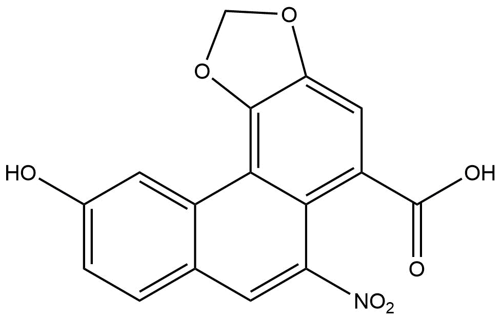


Product name: Aristolochic acid C
Synonym name: Aristolochic acid C
Catalogue No.: BPF0516
Cas No.: 4849-90-5
Formula: C16H9NO7
Mol Weight: 327.248
Botanical Source:
Physical Description:
Type of Compound: Alkaloids
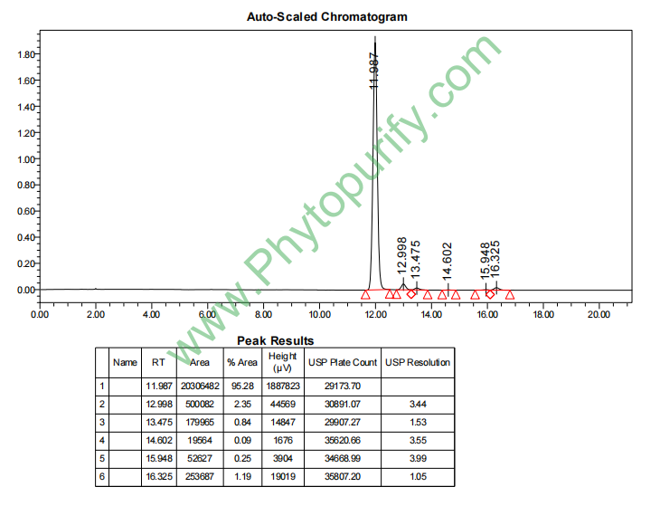
Purity: 95%~99%
Analysis Method: HPLC-DAD or/and HPLC-ELSD
Identification Method: Mass, NMR
Packing: Brown vial or HDPE plastic bottle
The product could be supplied from milligrams to grams
Inquire for bulk scale.
For Reference Standard and R&D, Not for Human Use Directly.
HPLC of Aristolochic acid C
Storage conditions:Short-term storage at 2~8℃, long-term storage at -20 ~ -80℃
119.1300
3.2289
.1387
.1729
1.8125
18.6614
Low
91.2244
2.6613
Yes
No
No
No
Yes
Yes
2.4
Yes
Yes
Yes
Yes
Aristolochic acids (AAs) are a class of naturally occurring compounds in the Aristolochia genus(Aristolochia)And Asarum genus(Asarum)Nitrophenanthrene carboxylic acid derivatives in plants have attracted much attention due to their significant nephrotoxicity and carcinogenicity. Since the first report of Aristolochic Acid Nephropathy (AAN) in the 1990s, this type of compound has become a hot topic in natural product toxicology research. Aristolochic acid C (AAC), as an important member of the Aristolochic acid family, shares similarities in chemical structure, biological activity, and toxicity mechanisms with aristolochic acid I (AAI) and aristolochic acid II (AAII), while also exhibiting unique molecular characteristics.
The CAS number of aristolochic acid C is 4849-90-5, and its molecular formula is C ₁₇ H ₁∝ NO ₆. It belongs to the class of nitrophenanthrene compounds. Compared with AAI, AAC lacks a methoxy group at the C-8 position of the phenanthrene ring, while retaining a hydroxyl group. This structural difference not only affects its physicochemical properties, but also has a profound impact on its interaction mode with biomolecules. Early research mainly focused on the toxicity of AAI and AAII, but with further exploration of aristolochic acid metabolomics and toxicology, the unique role of AAC in nephrotoxicity, oxidative stress, and cell apoptosis regulation has gradually been revealed.
It is worth noting that aristolochic acid C is not only an inhibitor of phospholipase A2 (PLA2), but also can disrupt the periplasmic microtubule array of Arabidopsis interphase cells and inhibit root growth, suggesting that it may exert biological effects by interfering with cytoskeleton dynamics. In mammalian systems, the nephrotoxicity of AAC involves BCL2 family proteins, NFE2L2 antioxidant pathway, TP53 tumor suppressor factor, and CASP3 mediated apoptosis cascade, which together constitute the complex pathological network of aristolochic acid nephropathy.
This article will provide a systematic review of the research progress of aristolochic acid C from the perspectives of chemical structure, plant origin, pharmacological activity, molecular mechanism, pharmacological evaluation, and clinical application prospects, aiming to provide reference for natural product toxicology and drug safety evaluation.
The molecular formula of aristolochic acid C is C ₁₇ H ₁∝ NO ₆, with a molecular weight of 327.2480 g/mol. Its core structure is a phenanthrene ring skeleton, with a carboxyl group (- COOH) connected to the C-1 position of the phenanthrene ring, a nitro group (- NO ₂) connected to the C-4 position, a hydroxyl group (- OH) at the C-8 position, and a methoxy group (- OCH ∝) at the C-10 position. Compared with aristolochic acid I (AAI, C ₁₇ H ₁₁ NO ₇, with methoxy group at C-8 position), AAC replaces methoxy group with hydroxyl group at C-8 position. This structural difference enhances its polarity and hydrogen bond donor ability. Compared with aristolochic acid II (AAII, C ₁₆ H ₉ NO ₆, with a hydrogen atom at the C-8 position), the hydroxyl group at the C-8 position of AAC gives it stronger hydrophilicity and reactivity.
According to computational chemistry predictions and experimental measurements, the physicochemical properties of aristolochic acid C are as follows:
The UV visible absorption spectrum of aristolochic acid C exhibits characteristic absorption peaks in the range of 250-400 nm, with strong absorption at approximately 260 nm and 320 nm attributed to the π →π of the phenanthrene ring Transition, and weak absorption at around 390 nm may be related to the n →π of nitro groups Transition related. In the infrared spectrum, the strong absorption peak at approximately 1700 cm ⁻¹ corresponds to the C=O stretching vibration of carboxyl groups, while the absorption peaks at approximately 1520 cm ⁻¹ and 1350 cm ⁻¹ correspond to the asymmetric and symmetric stretching vibrations of nitro groups, respectively. In the nuclear magnetic resonance hydrogen spectrum, the aromatic proton signal on the phenanthrene ring is distributed in the δ 7.0-8.5 ppm region, with the proton signal of the C-8 hydroxyl group appearing in the δ 9.5-10.5 ppm region and easily exchanging with the solvent.
Aristolochic acid C mainly exists in the Aristolochia genus(Aristolochia)And Asarum genus(Asarum)Among plants, these plants have a long history of application in traditional Chinese medicine, Ayurvedic medicine, and South American folk medicine. The following are representative plants with high AAC content:
The extraction of aristolochic acid C is usually carried out by combining organic solvent extraction with chromatographic separation. The specific process is as follows:
1. Raw material pretreatment Crush the dried plant material to 40-60 mesh, degrease it with petroleum ether or n-hexane, and remove chlorophyll and lipid soluble impurities.
2. Solvent extraction Using methanol or 70% ethanol aqueous solution as the extraction solvent, with a material to liquid ratio of 1:10-1:20 (w/v), reflux extraction is carried out 2-3 times at 60-80 ℃ for 2-4 hours each time. Ultrasound assisted extraction (40-60 kHz, 30-60 minutes) can improve extraction efficiency by about 20-30%.
3. Liquid liquid extraction After concentrating the extract, liquid-liquid extraction was performed using ethyl acetate or n-butanol, and the aristolochic acid compounds were mainly distributed to the ethyl acetate phase. Adjusting the pH to 2-3 (using dilute hydrochloric acid) can promote AAC to enter the organic phase in free acid form.
4. Column chromatography separation Using silica gel column chromatography (200-300 mesh) with chloroform methanol acetic acid (90:10:1 to 70:30:1) gradient elution, collect the fraction containing AAC. Further purification by Sephadex LH-20 gel column chromatography (methanol elution) or preparative high performance liquid chromatography (C18 reverse phase column, acetonitrile water formic acid system) can obtain AAC monomer with purity>98%.
5. Quality control Quantitative analysis is performed using high-performance liquid chromatography diode array detection (HPLC-DAD) or liquid chromatography-mass spectrometry (LC-MS/MS), with detection wavelengths typically set at 250 nm or 320 nm. The retention time of AAC is typically between AAII and AAI on a C18 column, consistent with the polarity effect of hydroxyl substituents.
The nephrotoxicity of aristolochic acid C is its most concerning pharmacological activity and the main obstacle limiting its medicinal value. Both in vivo and in vitro experiments have confirmed that AAC can induce damage to renal tubular epithelial cells, with a toxicity intensity of about 1/5-1/3 of AAI, but higher than AAII.
Cell level research In human renal proximal tubular epithelial cells (HK-2 cells), AAC (10-100 μ M) treatment for 24-48 hours can lead to a concentration dependent decrease in cell viability, with a half maximal inhibitory concentration (IC ₅₀) of approximately 40-60 μ M. Morphological observations showed that cells treated with AAC exhibited typical apoptotic features such as nuclear condensation, formation of apoptotic bodies, and mitochondrial swelling. Flow cytometry analysis showed that AAC can induce cell cycle arrest in the G0/G1 phase and increase the proportion of hypodiploid cells.
Animal model research In C57BL/6 mice, intraperitoneal injection of AAC (5-20 mg/kg/d, continuous for 7 days) can significantly increase blood urea nitrogen (BUN) and serum creatinine (SCr) levels, and renal histopathological examination shows degeneration, necrosis, and interstitial fibrosis of renal tubular epithelial cells. It is worth noting that there are significant gender differences in the nephrotoxicity of AAC, with male mice experiencing more severe kidney damage than females, which may be related to the regulation of metabolic enzyme activity by androgens.
Aristolochic acid C is a competitive inhibitor of phospholipase A2 (PLA2), with an IC ₅₀ of approximately 15-25 μ M. PLA2 catalyzes the hydrolysis of sn-2 fatty acids in membrane phospholipids, releasing arachidonic acid, which is a key enzyme in inflammatory reactions. AAC binds to the hydrophobic channel of PLA2 through its phenanthrene ring skeleton, while nitro and carboxyl groups form hydrogen bonds with histidine and aspartic acid residues at the active site. This inhibitory activity may partially explain the anti-inflammatory effects of aristolochic acid compounds, but it may also exacerbate cell damage by interfering with membrane phospholipid metabolism.
In Arabidopsis thaliana(Arabidopsis thaliana)In root tip cells, AAC (10-50 μ M) treatment can cause disruption of the periplasmic microtubule array in interphase cells, manifested as microtubule bundle breakage, depolymerization, and abnormal aggregation. This effect is not related to the direct binding of microtubules, but rather achieved through the activation of the phosphorylation signaling pathway of microtubule associated proteins. The difference in the destructive effect of AAC on plant microtubules and its impact on animal cell microtubules suggests that it may have species selective cytoskeletal toxicity.
The nephrotoxicity of aristolochic acid C involves cross regulation of multiple signaling pathways, among which BCL2 family proteins, NFE2L2 antioxidant pathway, TP53 tumor suppressor factor, and CASP3 mediated apoptosis cascade constitute the core mechanism network.
1. BCL2 family and mitochondrial apoptosis pathway
AAC treatment can lead to downregulation of anti apoptotic protein BCL2 expression and upregulation of pro apoptotic protein BAX expression in renal tubular epithelial cells, with a significant increase in BAX/BCL2 ratio. This change promotes the translocation of BAX from the cytoplasm to the outer membrane of mitochondria, forming oligomeric channels, leading to a decrease in mitochondrial membrane potential (Δ PSI m), release of cytochrome c into the cytoplasm, and activation of CASP9 and CASP3, ultimately executing the apoptotic program. Immunofluorescence staining showed that in HK-2 cells treated with AAC, the mitochondrial localization of BAX increased by about 3-5 times, while the mitochondrial protective effect of BCL2 was weakened.
2. NFE2L2 antioxidant pathway
NFE2L2 (NRF2) is the main transcription factor for cellular antioxidant defense. AAC can induce the dissociation and nuclear translocation of NFE2L2 from KEAP1 complex, upregulating the expression of downstream antioxidant enzymes such as heme oxygenase-1 (HO-1), NAD (P) H quinone oxidoreductase 1 (NQO1), and glutathione S-transferase (GST). However, this adaptive antioxidant response gradually exhausts under continuous exposure, leading to the accumulation of reactive oxygen species (ROS). It is worth noting that the ROS induced by AAC mainly comes from the activation of NADPH oxidase (NOX2), rather than the mitochondrial electron transport chain. NOX2 inhibitors (such as apocynin) can partially alleviate the cytotoxicity of AAC, indicating that oxidative stress plays a key role in renal injury.
3. TP53 signaling pathway
AAC can activate TP53 (p53) protein, manifested by increased phosphorylation (Ser15 site) and acetylation levels of p53, as well as upregulation of its transcription target genes CDKN1A (p21) and BAX. The induction of p21 leads to cell cycle arrest at the G1/S checkpoint, buying time for DNA damage repair. However, when DNA damage exceeds its repair capacity, p53 instead promotes apoptosis. In HK-2 cells with p53 knockout, the cytotoxicity of AAC was significantly reduced, indicating that p53 is a key mediator of AAC nephrotoxicity.
4. Renal injury markers
AAC treatment can induce significant upregulation of renal injury molecule-1 (KIM-1/HAVCR1) and neutrophil gelatinase associated lipocalin (NGAL/LCN2) expression. KIM-1 is a transmembrane glycoprotein that is highly expressed in damaged renal tubular epithelial cells and can serve as a biomarker for early renal injury. NGAL is involved in iron ion transport and inflammatory response, and its urinary excretion is positively correlated with the degree of renal tubular injury. In the mouse model treated with AAC, urinary KIM-1 and NGAL levels increased within 24 hours after administration, earlier than changes in blood creatinine and urea nitrogen.
The nitro group of aristolochic acid C is metabolized by reductases such as NAD (P) H quinone oxidoreductase and cytochrome P450 reductase in cells to form N-hydroxyaristolochic acid lactam, which further forms covalent adducts with adenine and guanine bases in DNA. The main types of AAC-DNA adducts are 7- (deoxyadenosine-N ⁶ - yl) aristolochic acid lactam (dA AAI) and 7- (deoxyguanosine-N ² - yl) aristolochic acid lactam (dG AAI). These adducts can cause DNA polymerase reading errors, leading to A → T transition mutations, which are the molecular basis for the carcinogenicity of aristolochic acid. Compared with AAI, AAC forms fewer DNA adducts, but the adducts have a longer retention time in tissues, which may be related to the metabolic stability of its C-8 hydroxyl group.
The effect of AAC on microtubules is not through direct binding to microtubule proteins, but through activation of the RhoA/ROCK signaling pathway, leading to excessive phosphorylation of microtubule associated protein MAP4. Phosphorylated MAP4 dissociates from the surface of microtubules, reducing their stability and ultimately leading to the depolymerization of microtubule skeletons. In addition, AAC can inhibit the localization of microtubule positive end tracking proteins (such as EB1), interfere with the interaction between microtubules and the cell cortex, and affect cell polarity and directional migration.
Based on Lipinski's Five Rules and Veber's Rules, the pharmacological parameters of Aristolochic Acid C are as follows:
From the above parameters, AAC basically meets the physical and chemical properties requirements of oral drugs, but a high TPSA may limit its intestinal absorption. In addition, a positive Ames test result suggests a genetic toxicity risk, which is the biggest obstacle to drug development.
absorb The oral bioavailability of AAC is relatively low (about 15-25% in rat experiments), which may be related to its limited solubility in the gastrointestinal tract and the efflux of P-glycoprotein (P-gp). The Caco-2 cell monolayer experiment showed that the apparent permeability coefficient (Papp) of AAC was 1.2-2.5 × 10 ⁻⁶ cm/s, indicating moderate permeability of the compound.
distribution AAC is widely distributed in the body, with the highest concentrations in the kidneys and liver, which is consistent with the target organs of nephrotoxicity and hepatotoxicity. The plasma protein binding rate is about 85-92%, mainly binding to albumin. The apparent volume of distribution (Vd) is approximately 0.5-1.0 L/kg, indicating limited tissue distribution.
Metabolism The main metabolic pathways of AAC include: ① reduction of nitro groups to N-hydroxyaristolochic acid lactam (catalyzed by cytochrome P450 1A2 and NAD (P) H quinone oxidoreductase); ② C-8 hydroxy glucuronic acid binding (catalyzed by UGT1A1 and UGT1A9); ③ Oxidation of phenanthrene ring (catalyzed by CYP1A2 and CYP3A4). Metabolites are mainly excreted through bile and partially filtered through the glomerulus.
excretion The half-life (t ₁/₂) of AAC and its metabolites is approximately 8-12 hours, but the clearance half-life of DNA adducts can reach several weeks to months. About 60-70% of the administered dose is excreted through feces within 72 hours, and 20-30% is excreted through urine. It is worth noting that the accumulation of AAC in the kidneys is an important cause of its long-term nephrotoxicity.
Based on existing data, the toxicity risk of aristolochic acid C can be summarized as follows:
Aristolochic acid C, as one of the active ingredients of Aristolochia plants, has been widely present in traditional Chinese medicine preparations, such as "Longdan Xiegan Wan" and "Bazheng San". However, since 2003, multiple countries and regions have banned or restricted the use of traditional Chinese medicinal materials containing aristolochic acid compounds. However, due to plant identification errors, improper use of substitutes, and illegal trade, aristolochic acid nephropathy still occurs from time to time worldwide.
Although nephrotoxicity limits the direct medicinal value of aristolochic acid C, its unique molecular structure provides insights for drug design:
Structural modification Reducing toxicity through chemical modification, such as reducing nitro groups to amino groups or esterifying carboxyl groups, may result in derivatives with lower toxicity. Previous studies have reported that the nephrotoxicity of AAC methyl ester, a derivative of AAC, is reduced by about 5 times, while still retaining PLA2 inhibitory activity.
Targeted delivery Targeted delivery of AAC to tumor tissues using nanocarriers such as liposomes and polymer micelles can reduce systemic exposure. Preliminary studies have shown that the nephrotoxicity of AAC liposomes in tumor bearing mice is reduced by about 60% compared to free drugs.
As a tool molecule AAC, as a PLA2 inhibitor and microtubule disruptor, can be used to study the molecular mechanisms of inflammation and cytoskeletal dynamics. Its unique ability to form DNA adducts also makes it an ideal model compound for studying chemical carcinogenesis mechanisms.
Considering the genetic and renal toxicity of aristolochic acid C, the following measures are recommended:
Aristolochic acid C, as an important member of the Aristolochic acid family, shares similarities in chemical structure, pharmacological activity, and toxicity mechanism with AAI and AAII, while exhibiting unique molecular characteristics. The introduction of the C-8 hydroxyl group alters its polarity and metabolic pathway, resulting in a renal toxicity intensity lower than AAI but higher than AAII. AAC induces damage to renal tubular epithelial cells through the BCL2/BAX mediated mitochondrial apoptosis pathway, NFE2L2 antioxidant pathway, TP53 signaling pathway, and NOX2 oxidative stress pathway, and its ability to form DNA adducts constitutes the basis of genetic toxicity.
Although the direct medicinal value of AAC is limited by nephrotoxicity, its activity as a PLA2 inhibitor and microtubule disruptor provides a structural template for drug design. Through chemical modification, targeted delivery, and toxicity avoidance strategies, it is expected to develop low toxicity derivatives. Meanwhile, AAC, as a model compound for studying the mechanisms of chemical carcinogenesis and nephrotoxicity, has significant value in basic toxicology research.
In the future, with the development of metabolomics, epigenetics, and single-cell sequencing technologies, the understanding of the toxicity mechanism of aristolochic acid C will be further deepened. Establishing regulatory standards based on risk assessment, strengthening the safety evaluation of traditional Chinese medicine, and developing effective detoxification strategies are key measures to safeguard public health. The research process of aristolochic acid C warns us that natural products are not only treasure trove for drug discovery, but also may conceal potential health risks. Only through rigorous scientific evaluation can traditional medicine be modernized and sustainably developed.
