


Product name: Lysophosphatidylcholine
Synonym name: 1-Stearoyl-sn-glycero-3-phosphocholine;1-Stearoyl-2-hydroxy-sn-glycero-3-phosphocholine
Catalogue No.: BP2371
Cas No.: 19420-57-6
Formula: C26H54NO7P
Mol Weight: 523.692
Botanical Source:
Type of Compound: Aliphatics
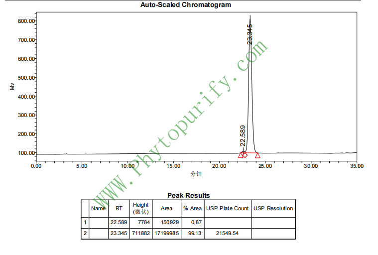
Purity: 95%~99%
Analysis Method: HPLC-DAD or/and HPLC-ELSD
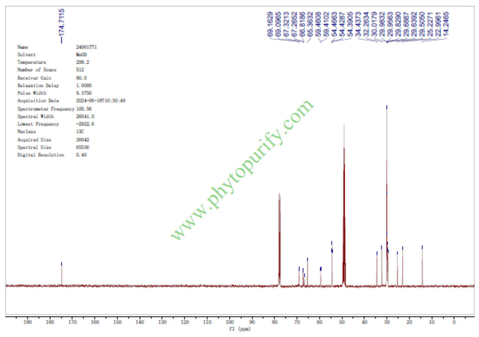
Identification Method: Mass, NMR
Packing: Brown vial or HDPE plastic bottle
Storage: Store in a well closed container, protected from air and light. Put into refrigerate or freeze for long term storage.
Whenever possible, you should prepare and use solutions on the same day. However, if you need to make up stock solutions in advance, we recommend that you store the solution as aliquots in tightly sealed vials at -20℃. Generally, these will be useable for up to two weeks.
The product could be supplied from milligrams to grams
Inquire for bulk scale.
For Reference Standard and R&D, Not for Human Use Directly.
HPLC of Lysophosphatidylcholine
NMR of Lysophosphatidylcholine
Storage conditions:Short-term storage at 2~8℃, long-term storage at -20 ~ -80℃
102.2900
2.6409
2.5626
.1652
.2811
.1776
Low
76.7759
3.7483
No
No
No
No
No
No
0.3
No
No
No
No
Lysophosphatidylcholine (LPC) is a class of bioactive lipid molecules produced by the hydrolysis of phosphatidylcholine (PC) catalyzed by phospholipase A2 (PLA2). Its structural feature is that the sn-1 or sn-2 position of the glycerol skeleton contains only one fatty acid chain, while the other position is a hydroxyl group, and the phosphocholine group is connected to the sn-3 position. According to the length, saturation, and position (sn-1 or sn-2) of the fatty acid chain, LPC exists in various molecular species, among which 1-stearoyl-sn-glycerin-3-phosphocholine (CAS number: 19420-57-6) is LPC 18:0, representing a specific subtype of sn-1 linked stearoyl (C18:0).
For a long time, LPC has been regarded as an intermediate or "junk" molecule in cell membrane phospholipid metabolism, but research in the past two decades has completely overturned this cognition. LPC has been proven to be a multifunctional signaling molecule widely involved in physiological and pathological processes such as cell proliferation, migration, inflammatory response, oxidative stress, and cell apoptosis. Its biological effects are mainly achieved through the activation of G protein coupled receptors (GPCRs) such as G2A, GPR4, and GPR119, as well as through non receptor dependent pathways such as insertion into the cell membrane to alter membrane fluidity. In pathological conditions, especially in inflammation and autoimmune diseases, the local concentration of LPC significantly increases, becoming one of the key driving factors for disease progression.
Of particular note is the increasingly prominent role of LPC in demyelinating diseases of the central nervous system (CNS). Demyelinating diseases, such as multiple sclerosis (MS), are characterized by the destruction of myelin sheaths and damage to oligodendrocytes. Both clinical and basic research have shown that LPC levels are significantly elevated in the cerebrospinal fluid and lesion tissues of MS patients. Exogenous LPC local injection has been widely used as a standard method for establishing experimental demyelination animal models, such as LPC induced demyelination models. LPC initiates and exacerbates the demyelination process by inducing inflammatory responses, activating astrocytes and microglia, directly damaging oligodendrocytes, and disrupting the integrity of the blood-brain barrier (BBB). Therefore, a deep understanding of the pharmacological properties of LPC and its molecular mechanisms in demyelinating diseases is of great significance for developing new therapeutic strategies.
This article will provide a systematic review of LPC (especially 1-stearoyl-sn-glycerin-3-phosphocholine) from the aspects of chemical structure, physicochemical properties, pharmacological activity, mechanism of action, drug evaluation, and clinical application prospects, aiming to provide reference for natural product pharmacology and neurodegenerative disease research.
The chemical structure of 1-stearoyl-sn-glycerin-3-phosphocholine exhibits typical LPC characteristics. The sn-1 position of its glycerol skeleton is connected by an ester bond to a stearoyl group (C18:0), which is a saturated octadecan fatty acid; Sn-2 position is a free hydroxyl group; The sn-3 position is connected to a phosphocholine group. Its molecular formula is C ₂₆ H ₅₄ NO ₇ P, and its molecular weight is 524.70 g/mol. This structure endows the molecule with amphiphilicity: the tail of long-chain fatty acids is a hydrophobic region, while the head of phosphatidylcholine and free hydroxyl groups are hydrophilic regions. This amphiphilicity is the structural basis for its formation of micelles, interaction with cell membranes, and as a signaling molecule.
According to the calculated chemical parameters, the lipid water partition coefficient (LogP) of LPC 18:0 is 2.64, indicating its moderate lipophilicity and ease of insertion into lipid bilayers. The topological polar surface area (TPSA) is 102.29 Å ², indicating that it has a certain polarity, but not enough to freely penetrate the cell membrane. Low water solubility (0.1652 mg/mL), tends to form micelles or bind to carrier proteins (such as albumin) in physiological solutions.
In terms of drug properties, the molecular weight of LPC 18:0 (524.70 Da) is slightly higher than the traditional Lipinski's Rule of Five limit of molecular weight<500, but its LogP value (2.64) and the number of hydrogen bond donors/acceptors (3 and 8, respectively) comply with the rules. It is worth noting that its blood-brain barrier (BBB) penetration ability is evaluated as "low", which is related to its polar head and relatively large molecular weight. However, under pathological conditions such as inflammation and ischemia, BBB integrity is impaired, and LPC can still enter the CNS through passive diffusion or carrier mediated transport. In addition, hERG inhibition prediction is negative, indicating a low risk of cardiac toxicity; The Ames test result is 0.3, indicating a low risk of genetic toxicity. These preliminary pharmacological evaluations provide a basis for the feasibility of using it as a drug lead or target for research.
Although LPC is widely present in organisms, as a natural product, it is not a secondary metabolite directly synthesized and accumulated in large quantities by plants. On the contrary, LPC is a product of phospholipid metabolism in animal and plant cell membranes. In plants, LPC mainly exists as an intermediate in phospholipid metabolism and can be detected in tissues such as seeds, leaves, and roots. For example, in the seeds of oil crops such as soybeans, rapeseed, and sunflowers, due to their high content of phospholipids, the activation of endogenous PLA2 during seed germination or processing can lead to the production of LPC. In addition, extracts from certain medicinal plants such as ginkgo and ginseng also contain trace amounts of LPC, but their content is usually much lower than the main active ingredients.
Due to the low and unstable content of LPC in natural plants, its acquisition usually does not rely on direct plant extraction, but rather on chemical synthesis or enzymatic synthesis. However, the preparation of LPC from natural phospholipid resources still has research value, and the main methods include:
Solvent extraction and fractionation Starting from raw materials rich in phospholipids (such as soy lecithin), total lipid extraction is carried out using solvents such as ethanol and chloroform methanol. Subsequently, LPC components can be preliminarily separated by silica gel column chromatography and gradient elution using different solvent systems (such as chloroform methanol water). This method is easy to operate, but has low purity and yield, and is prone to introducing other lipid impurities.
Enzymatic hydrolysis This is currently the most commonly used method for preparing specific molecular species LPC. Using high-purity phospholipase A2 (such as PLA2 from snake venom or pig pancreas) to hydrolyze natural PC under specific conditions (such as pH 8.0, containing Ca ² ⁺). PLA2 specifically hydrolyzes sn-2 fatty acids, generating LPC and free fatty acids of sn-1 acyl groups. After the reaction, the target LPC is isolated and purified by solvent extraction or chromatography (such as high-performance liquid chromatography, HPLC). This method has mild reaction conditions, high selectivity, and can obtain high-purity single molecular species LPC (such as LPC 18:0).
Chemical Synthesis For LPC with clear structure, such as 1-stearoyl-sn-glycerin-3-phosphocholine, chemical total synthesis is a reliable way to obtain high-purity and structurally determined products. Typically, chiral glycerol derivatives (such as (R) - glycidol) are used as starting materials, and a protective group strategy is employed to sequentially introduce stearoyl and phosphocholine groups. This method is cumbersome in steps and requires high synthesis technology, but it can accurately control the position and stereoconfiguration of fatty acid chains.
In practical research, LPC 18:0 used for pharmacological experiments is usually purchased directly from commercial suppliers such as Avanti Polar Lipids, with a purity of over 99%. For the study of plant derived LPC, the focus is on analyzing its content changes in specific plant tissues and its relationship with plant physiological processes such as stress resistance and signal transduction, rather than extracting it as the main active ingredient.
LPC has a wide range of pharmacological activities, involving multiple systems, particularly in inflammation, immune regulation, and the nervous system. The activity research of demyelinating diseases is particularly in-depth.
LPC is widely recognized as a potent pro-inflammatory mediator. LPC can induce the expression of pro-inflammatory cytokines (such as TNF - α, IL-6, IL-1 β) and chemokines in various cell types, such as endothelial cells, smooth muscle cells, and macrophages. In the demyelinating disease model, direct injection of LPC into the white matter of the brain can rapidly activate astrocytes (manifested as upregulation of GFAP expression) and microglia (increased Iba-1 positive cells), triggering a strong local inflammatory response. This inflammatory response is the direct cause of subsequent damage to oligodendrocytes and demyelination. Research has shown that LPC induced release of TNF - α and IL-6 can further amplify the inflammatory cascade by activating the NF - κ B and MAPK signaling pathways, forming a vicious cycle.
LPC has clear cytotoxicity and can induce apoptosis in various cells. In oligodendrocyte lines such as OLN-93, LPC treatment can lead to a decrease in mitochondrial membrane potential, release of cytochrome c, activation of caspase-3, and ultimately induce apoptosis. This process is closely related to LPC induced oxidative stress (ROS production) and calcium overload. LPC 18:0, as an inducer of cell apoptosis, partially relies on its insertion into the cell membrane to alter the physical properties of the membrane (such as increasing membrane fluidity and promoting lipid raft formation), thereby affecting the functions of membrane receptors and signaling proteins. In the demyelination model, the direct killing effect of LPC on oligodendrocytes is one of the core mechanisms leading to myelin sheath disintegration.
The integrity of BBB is crucial for maintaining CNS homeostasis. LPC can significantly impair BBB function. In vitro experiments have shown that LPC can reduce the transendothelial resistance (TEER) of brain microvascular endothelial cells and increase cell paracellular permeability. The mechanism involves downregulating the expression of tight junction proteins such as Claudin-5 and Occludin, as well as activating the RhoA/ROCK signaling pathway in endothelial cells, leading to cytoskeleton rearrangement. In the body, BBB leakage can be observed early after LPC injection, making it easier for peripheral immune cells (such as T cells and macrophages) and inflammatory factors to enter the CNS, thereby exacerbating demyelinating lesions.
LPC treatment can directly affect the structural proteins of myelin sheaths. In the demyelination model, the immunohistochemical staining intensity of MBP (myelin basic protein) and PLP (proteolipid protein) significantly decreased, indicating the destruction of myelin sheath structure. LPC not only indirectly reduces MBP and PLP by damaging oligodendrocytes, but may also directly degrade myelin proteins by activating matrix metalloproteinases (MMPs). In addition, the inflammatory environment induced by LPC can inhibit the differentiation and maturation of oligodendrocyte precursor cells (OPCs), hindering the regeneration and repair of myelin sheaths.
The biological effects of LPC are mediated through multiple mechanisms and molecular targets, and its complexity determines its multiple roles in diseases.
LPC is an endogenous ligand for various GPCRs, among which G2A (GPR132) and GPR4 are the most extensively studied.
- G2A receptor LPC is an agonist of G2A. After G2A activation, it can couple with G α i and G α q proteins, thereby regulating downstream signaling pathways such as inhibiting cAMP production, activating the PLC - β/IP3/Ca ² ⁺ pathway, and MAPK (ERK, p38, JNK) pathway. In immune cells, G2A mediates LPC induced chemotaxis and cytokine release. In the demyelination model, G2A is upregulated in astrocytes and microglia, and its activation is a key link in LPC induced neuroinflammation.
- GPR4 receptor LPC can also activate GPR4, mainly coupling G α s and G α q. Activation of GPR4 can increase cAMP levels and promote cell migration. GPR4 is highly expressed in endothelial cells, and LPC mediates the disruption of endothelial barrier function and angiogenesis through GPR4.
- GPR119 receptor LPC is an agonist of GPR119, which is mainly expressed in pancreatic beta cells and intestinal L cells, and is involved in regulating the secretion of insulin and incretin (such as GLP-1). This pathway is related to metabolic regulation, but its function in the CNS is not yet clear.
In addition to GPCR, LPC can also exert its effects through non receptor pathways:
- Membrane fluidity regulation LPC, as an amphiphilic molecule, can be inserted into the lipid bilayer of cell membranes to alter the physical properties of the membrane, such as increasing membrane fluidity and promoting the formation of non bilayer structures. This will directly affect the conformation and function of membrane proteins (such as ion channels, transporters, receptors), as well as the membrane localization of signaling molecules.
- Oxidative stress induction LPC can stimulate cells to produce a large amount of reactive oxygen species (ROS), leading to lipid peroxidation, DNA damage, and protein oxidation. Oxidative stress is an important upstream event in LPC induced cell apoptosis and inflammation. The mechanism may involve activation of NADPH oxidase (NOX) and damage to the mitochondrial electron transport chain.
- Calcium ion homeostasis disorder LPC can activate the PLC/IP3 pathway or directly act on calcium channels on the membrane, leading to an increase in intracellular calcium ion concentration. Calcium overload can activate various enzymes such as calpain and nitric oxide synthase (NOS), ultimately leading to cell damage.
In demyelinating diseases, the role of LPC is a complex network of multiple targets and pathways:
1. Direct damage LPC induces oxidative stress and calcium overload by inserting into the membrane of oligodendrocytes, activating the caspase cascade reaction and directly leading to apoptosis of oligodendrocytes.
2. Inflammation amplification LPC activates G2A receptors on astrocytes and microglia, activates NF - κ B and MAPK pathways, and leads to the massive release of pro-inflammatory factors such as TNF - α and IL-6. These cytokines, in turn, further activate glial cells, forming a positive feedback loop of inflammation.
3. BBB destruction LPC downregulates endothelial cell tight junction protein through GPR4 and RhoA/ROCK pathways, increases BBB permeability, and allows peripheral immune cells and inflammatory mediators to enter the CNS.
4. Myelin degradation LPC induced inflammatory environment activates MMPs, directly degrading myelin proteins such as MBP and PLP. Meanwhile, inflammatory factors inhibit the proliferation and differentiation of OPCs, hindering myelin regeneration.
5. Target association LPC directly or indirectly affects molecular targets closely related to demyelinating diseases through the above mechanisms:TNF and IL6 As a key pro-inflammatory factor, it is the core of the inflammatory cascade reaction;GFAP As a marker of astrocyte activation, it reflects the glial response induced by LPC;MBP and PLP As a structural protein of myelin sheath, its expression and integrity are direct indicators for measuring the degree of demyelination.
Based on the aforementioned physicochemical parameters, the pharmacological properties of LPC 18:0 exhibit duality.
- Advantage Moderate molecular weight, reasonable LogP value, and no risk of hERG inhibition or Ames toxicity suggest that it has a certain safety basis as a drug lead. Its clear pharmacological activity, especially in inducing demyelination, makes it an ideal tool compound for studying the mechanisms of demyelinating diseases and screening therapeutic drugs.
- challenge Its biggest challenge lies in Low blood-brain barrier penetration As a drug for treating CNS diseases, it needs to effectively enter the brain parenchyma. The low BBB penetration of LPC limits its potential as a systemic therapeutic drug. In addition, it Amphiphilia and metabolic instability It's also a problem. LPC can be rapidly hydrolyzed into lysophosphatidic acid (LPA) by hemolytic phospholipase D (autotaxin) in the blood, or further hydrolyzed into glycerophosphocholine by hemolytic phospholipase A1/A2. This rapid metabolism results in a short half-life and low bioavailability. Meanwhile, its pro-inflammatory activity may cause widespread side effects when administered systemically.
Given the pharmaceutical challenges of LPC, the possibility of developing it directly as a therapeutic drug (such as for promoting myelin regeneration) is low. On the contrary, its value is mainly reflected in the following aspects:
1. Disease modeling tool LPC is the gold standard tool for establishing demyelinating animal models, used to study disease mechanisms and screen candidate drugs.
2. Target discovery By studying the mechanism of action of LPC, new drug targets can be identified, such as G2A, GPR4, autotaxin, PLA2, etc. Inhibitors or antagonists targeting these targets may become a new strategy for treating demyelinating diseases.
3. Prodrug design Modifying the LPC structure, such as changing the length of fatty acid chains or introducing protective groups, to improve its metabolic stability and BBB penetration, may yield analogs with therapeutic potential.
At present, the direct application of LPC in clinical practice is very limited, mainly limited to research field As an inducer of demyelination models, it is widely used in neuroscience and pharmacology research. In addition, changes in plasma LPC levels have been used as biomarkers for certain diseases (such as atherosclerosis, diabetes, and Alzheimer's disease).
Lysophosphatidylcholine, especially 1-stearoyl-sn-glycerin-3-phosphocholine (LPC 18:0), has evolved from a neglected intermediate in phospholipid metabolism to a highly regarded bioactive molecule in the fields of natural product pharmacology and neuroscience. Its structure is simple, but it plays a key role in cell signal transduction, inflammation regulation, and cell fate determination through GPCR and non receptor pathways. In demyelinating diseases, LPC becomes the core driving force for disease initiation and progression by inducing inflammation, directly damaging oligodendrocytes, and disrupting the blood-brain barrier.
Although LPC itself has limited prospects as a therapeutic drug due to metabolic instability, low BBB penetration, and pro-inflammatory side effects, its value as a disease modeling tool and drug target discovery platform is irreplaceable. A deep understanding of the LPC signaling pathway has spurred the development of new drug pipelines targeting Autotaxin, G2A, GPR4, and other targets. In the future, with further elucidation of the complex network of action of LPC in CNS, as well as advances in drug chemistry and nanodelivery technology, we have reason to believe that a new generation of therapeutic strategies based on LPC biology will bring new hope to patients with demyelinating diseases. The study of LPC not only deepens our understanding of the role of lipid signaling in physiology and pathology, but also demonstrates a scientific discovery paradigm from basic metabolites to key disease regulatory factors.
